Understanding diabetic retinopathy
How diabetes can affect retinal blood vessels and why regular eye examinations matter.
AI-Powered Retinal Analysis
Upload a retinal image for research predictions from your Roboflow model, or run the local simulated demo. Neither replaces professional eye-care evaluation.
Roboflow: diabetic-retinopathy-screening-ai/1 · Research use only
Example Image
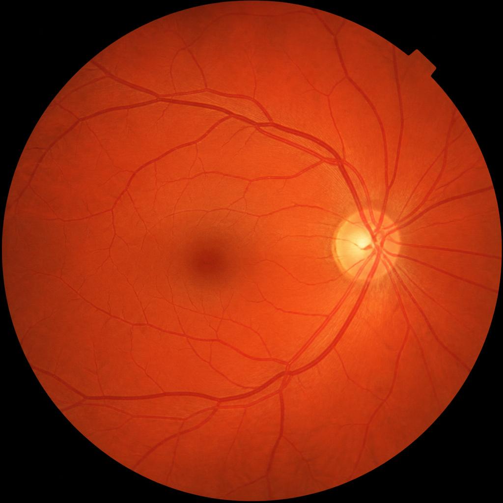
For best results
Use high-quality retinal fundus images with good focus, appropriate lighting, and the complete retinal area visible.
Process
Upload a retinal fundus image in JPG or PNG format.
EYESPY analyzes the image for visual features that may be associated with Diabetic Retinopathy.
The connected model returns classification scores across diabetic-retinopathy severity categories for the whole image. It does not locate individual lesions.
Receive a screening summary showing the model scores. These are model outputs, not clinically validated diagnostic probabilities.
Screening features
EYESPY is designed to look for retinal features that are commonly evaluated when screening for Diabetic Retinopathy. These are visual screening features only — the system does not independently provide a medical diagnosis, and no single feature is treated as proof of disease.
Small abnormalities involving retinal blood vessels.
Areas of bleeding visible within the retina.
Yellowish deposits that can occur in the retina.
Small areas associated with retinal nerve fiber layer abnormalities.
Abnormal new blood vessel growth that may occur in advanced retinal disease.
Candidate features are weighed together with image quality and how widely they are distributed. Isolated or ambiguous findings return an inconclusive result rather than a screening-positive one.
Resources
How diabetes can affect retinal blood vessels and why regular eye examinations matter.
Focus, lighting, centring and field of view guidance for usable screening images.
What a screening aid can and cannot tell you, and when to seek professional evaluation.
FAQ
Privacy
When live AI analysis is enabled and you provide consent, your uploaded retinal image is transmitted securely from EYESPY’s backend to Roboflow for AI inference. The image is therefore not processed exclusively within your browser. Please avoid uploading personally identifiable information. EYESPY is a research prototype and is not a substitute for professional medical evaluation.
Verified contact details have not been published yet.